Home
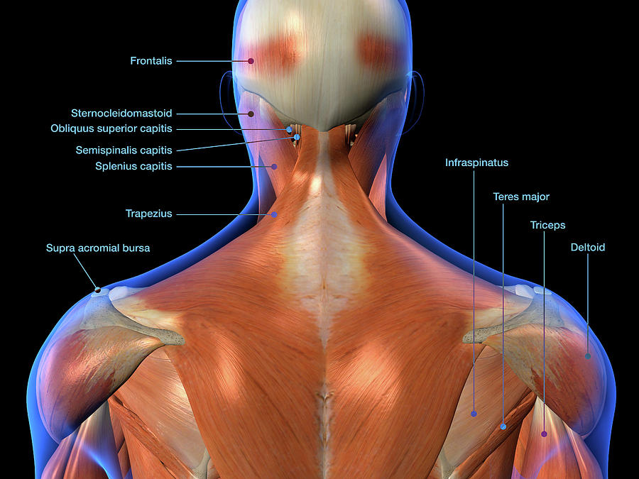
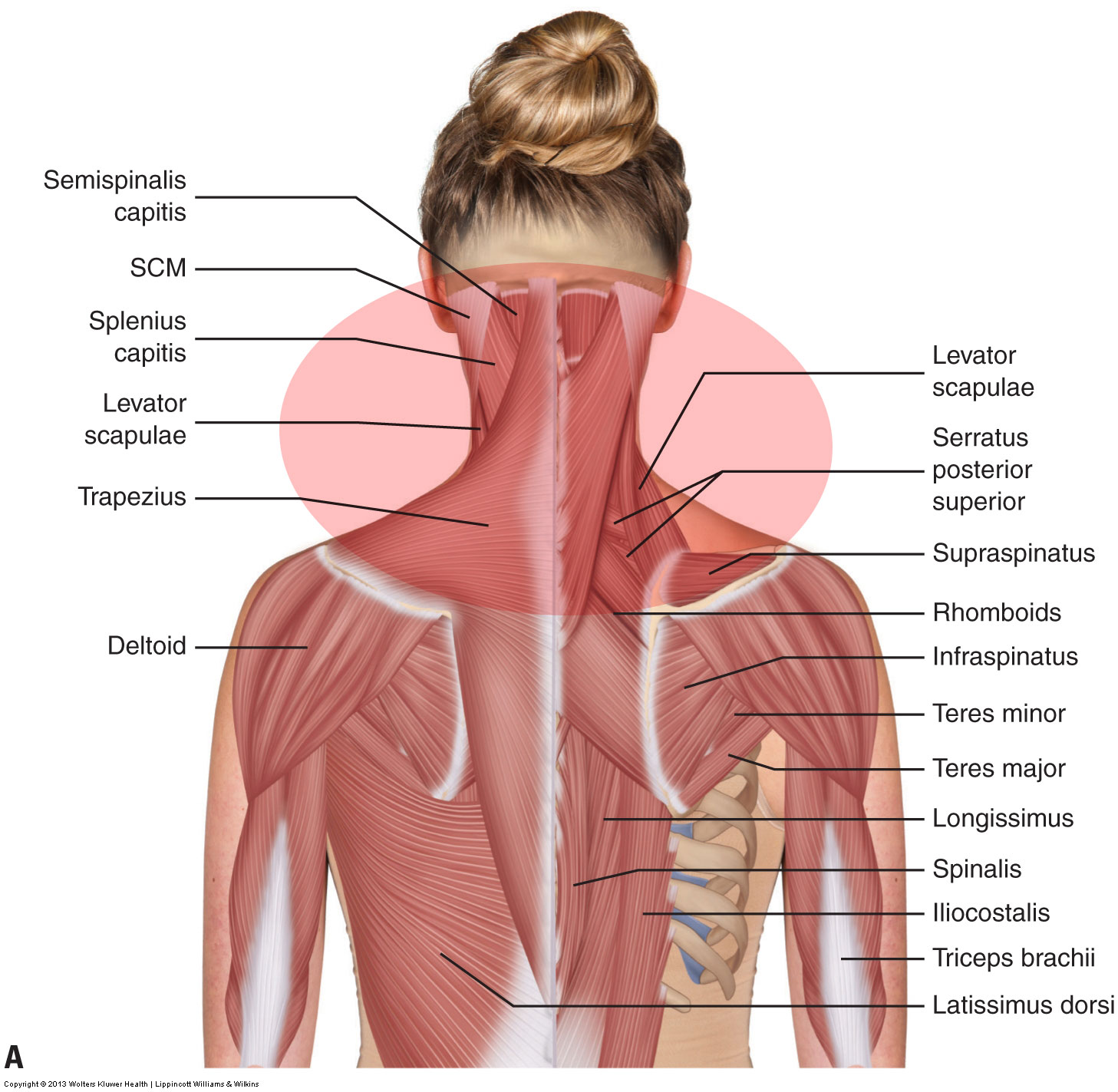
/ Anatomy Of The Back Of The Neck Muscles - Beautiful illustration of the deep and superficial ... : The image below to shows all the major back muscles (as well as some neck muscles):
Anatomy Of The Back Of The Neck Muscles - Beautiful illustration of the deep and superficial ... : The image below to shows all the major back muscles (as well as some neck muscles):
Anatomy Of The Back Of The Neck Muscles - Beautiful illustration of the deep and superficial ... : The image below to shows all the major back muscles (as well as some neck muscles):. Muscle spasming of the neck is likely the most common musculoskeletal complaint that exists. The superficial back muscles are covered by skin, subcutaneous connective tissue and a layer of fat. Four groups of muscles in neck. They move the head in every direction, pulling the skull and jaw towards the shoulders, spine, and scapula. Beneath the integument the back of neck presents in the median plane the ligamentum nuchae, which is a triangular fibrous sheet and represents upward continuation of supraspinous ligament.
In fact, the back contains a group of muscles, not one muscle. Intermediate back muscles and c. Watch cervical muscle anatomy animation. The extensors and rotators of the head and neck: Rectus capitis posterior major origin a…
Superficial And Deep Muscles Of The Neck Anatomy PNG Image ... from www.seekpng.com The splenius capitis and cervicis (spinotransversales muscles). If we want to locate the back muscles in the body, we can say that it starts from the top of the neck and ends. From the sides and the back of the neck, the splenius capitis inserts onto the head region, and the splenius. Its attachment to the frontal and occipital bellies (muscles on the brow at the front and on the upper back of the head). The muscles in all of the layers are innervated by the posterior rami of spinal nerves: Rectus capitis posterior major and rectus capitis posterior minor attach the inferior nuchal line of the occiput to the c2 and c1 vertebrae respectively. Several other muscles of the back also extend up to the neck region and are partly connected with the cervical part of the vertebral column, including the trapezius, levator scapulae, splenius, iliocostalis, longissimus, rotatores, semispinalis, interspinales, and intertransversarii muscles. The back anatomy includes some of the most massive and functionally important muscles in the human body.
Instead, i think it's simply better to break movements down into three categories
Beneath the integument the back of neck presents in the median plane the ligamentum nuchae, which is a triangular fibrous sheet and represents upward continuation of supraspinous ligament. The splenius capitis and cervicis (spinotransversales muscles). The back muscles can be three types. Explanations spinoscapular and spinohumeral muscles the extrinsic back muscles are also referred to as secondary back muscles. The splenius muscles originate at the midline and run laterally and superiorly to their insertions. The extensors and rotators of the head and neck: Muscles of the back can be divided into superficial, intermediate, and deep group. You can protect the back muscles by bending from the hip and knee when you lift objects from the ground. Other muscles in the back are associated with the movement of the neck and shoulders. Muscles of the head & neck | anatomy model. The muscles in all of the layers are innervated by the posterior rami of spinal nerves: Educational video describing the muscle anatomy of the neck. Rectus capitis posterior major and rectus capitis posterior minor attach the inferior nuchal line of the occiput to the c2 and c1 vertebrae respectively.
The physicians originally studying human anatomy thought the skull looked like an apple. Accordingly, the anterior (front) neck muscles can become long and weak. The platysma subcutaneous muscle of the neck (platysma) extends from the chin to the pectoral region. Obliquus capitis superior also extends from the occiput to c1 while obliquus. The superficial back muscles are covered by skin, subcutaneous connective tissue and a layer of fat.
Labeled Anatomy Chart Of Neck And Back Photograph by Hank ... from images.fineartamerica.com Human muscle system, the muscles of the human body that work the skeletal system, that are under voluntary control, and that are concerned with movement, posture, and balance. Table of contents the extrinsic muscles of the back: The muscles in all of the layers are innervated by the posterior rami of spinal nerves: The splenius muscles originate at the midline and run laterally and superiorly to their insertions. From the sides and the back of the neck, the splenius capitis inserts onto the head region, and the splenius. There are a number of specific muscles within the back anatomy, and its important to take a quick look at all of them to see how you can target them efficiently and develop a strong back. Injuries of the intrinsic back muscles often occur while using improper lifting technique. The splenius muscles originate at the midline and run laterally and superiorly to their insertions.
Table of contents the extrinsic muscles of the back:
The trapezius muscle can be involved in extending the head upward or neck backward. The physicians originally studying human anatomy thought the skull looked like an helmet. From the sides and the back of the neck, the splenius capitis inserts onto the head region, and the splenius. Instead, i think it's simply better to break movements down into three categories Obliquus capitis superior also extends from the occiput to c1 while obliquus. Muscle spasming of the neck is likely the most common musculoskeletal complaint that exists. There are a number of specific muscles within the back anatomy, and its important to take a quick look at all of them to see how you can target them efficiently and develop a strong back. The image below to shows all the significant back muscles (in addition to some neck muscles) The extensors and rotators of the head and neck: Working in pairs on the. Memorize all the muscle facts with the help of muscle cheat sheets. The suboccipital muscles act to rotate the head and extend the neck. Four groups of muscles in neck.
Instead, i think it's simply better to break movements down into three categories And the activity at our anterior neck muscles is connected in a wonderful way to the activity of. Memorize all the muscle facts with the help of muscle cheat sheets. Beneath the integument the back of neck presents in the median plane the ligamentum nuchae, which is a triangular fibrous sheet and represents upward continuation of supraspinous ligament. The image below to shows all the significant back muscles (in addition to some neck muscles)
What are the causes of muscle spasming in the neck? from learnmuscles.com The splenius muscles originate at the midline and run laterally and superiorly to their insertions. It arises from the oblique line of the lamina of thyroid cartilage. Since the all the back muscles originate in embryo (fetus) form by locations… they consist of: Muscles of the head & neck | anatomy model. The muscles in all of the layers are innervated by the posterior rami of spinal nerves: Several other muscles of the back also extend up to the neck region and are partly connected with the cervical part of the vertebral column, including the trapezius, levator scapulae, splenius, iliocostalis, longissimus, rotatores, semispinalis, interspinales, and intertransversarii muscles. These muscles give height and breadth to back development. Beneath the integument the back of neck presents in the median plane the ligamentum nuchae, which is a triangular fibrous sheet and represents upward continuation of supraspinous ligament.
Injuries of the intrinsic back muscles often occur while using improper lifting technique.
There are a number of specific muscles within the back anatomy, and its important to take a quick look at all of them to see how you can target them efficiently and develop a strong back. In fact, the back contains a group of muscles, not one muscle. The suboccipital muscles act to rotate the head and extend the neck. They move the head in every direction, pulling the skull and jaw towards the shoulders, spine, and scapula. Click now to learn more at kenhub! The thyrohyoid is a quadrilateral muscle located in the muscular triangle of the neck. Muscles of the head & neck | anatomy model. The splenius capitis and cervicis (spinotransversales muscles). To borrow a common phrase of renowned author and. Muscles of the back can be divided into superficial, intermediate, and deep group. Tutorials and quizzes on the anatomy and actions of the back muscles (iliocostalis, longissimus, spinalis, multifidus, and quadratus lumborum), using interactive animations, diagrams, and illustrations. This article provides an overview of the neck muscles, their anatomy, origins, insertions, actions, and innervation. Educational video describing the muscle anatomy of the neck.
This article covers the anatomy of the superficial muscles of the back, including trapezius, latissimus dorsi, levator scapulae, rhomboid major and minor anatomy of back of neck. Other muscles in the back are associated with the movement of the neck and shoulders.